Explore how PRP injections can treat morton's neuroma. Learn about platelet-rich plasma therapy benefits, recovery, and results for morton's neuroma.
Morton's neuroma, a painful thickening of the nerve tissue in the forefoot, is typically managed with corticosteroid injections as a first-line injectable treatment. PRP (Platelet-Rich Plasma) offers a biologically distinct alternative, and emerging evidence suggests it may produce more durable pain relief. For patients with chronic forefoot pain who have had limited or short-lived benefit from cortisone, PRP delivered under ultrasound guidance directly to the affected interdigital nerve is an increasingly used option before considering surgical neurectomy.
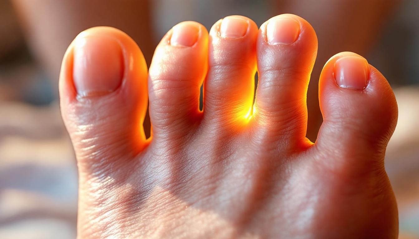
Morton's neuroma anatomy in the forefoot
What Is Morton's Neuroma?
Morton's neuroma is not a true tumor but a perineural fibrosis, a thickening of the fibrous tissue surrounding a plantar digital nerve, most commonly in the third intermetatarsal space (between the 3rd and 4th metatarsal heads), though the second space is also affected. The nerve becomes compressed and irritated by the surrounding metatarsal bones and the deep transverse metatarsal ligament, leading to progressive nerve thickening and degenerative changes.
Symptoms are characteristic: a burning, sharp, or shooting pain in the ball of the foot, often described as a sensation of standing on a pebble or a bunched-up sock. Numbness and tingling extending into the toes are common. Symptoms are typically worsened by tight footwear, high heels, or prolonged standing and walking, and are often relieved by removing shoes and massaging the foot. The Mulder's click, a palpable or audible click produced by squeezing the metatarsals together, is a positive clinical sign on examination.
Ultrasound or MRI confirms the diagnosis by identifying the hypoechoic nodule within the intermetatarsal space. Conservative management begins with footwear modifications (wide toe box, metatarsal pads, orthotics) and activity modifications. When conservative measures fail, corticosteroid injections are the standard first-line injectable treatment. For patients who fail multiple cortisone injections, options escalate to alcohol sclerosing injections, PRP, radiofrequency ablation, or surgical neurectomy.
Application
Patient Profile
Evidence Level
Typical Protocol
Best Candidates
Interdigital Neuroma Relief
Patients with confirmed Morton's neuroma on ultrasound, failed conservative care 3+ months
Level II–III (case series, small RCTs)
1–2 ultrasound-guided injections, 4–6 weeks apart
Single interspace neuroma, no surgical history, pain 4+ on VAS
Post-Surgical Neuroma Pain
Patients with residual or recurrent pain after prior excision or nerve surgery
Level III (clinical consensus, case reports)
2–3 injections spaced 4–6 weeks, combined with offloading
Recurrent neuroma confirmed on MRI/ultrasound, pain >6 months post-op
How PRP Works for Morton's Neuroma
The pathology of Morton's neuroma is degenerative, involving perineural fibrosis, nerve compression, and chronic low-grade inflammation rather than pure acute inflammation. This makes corticosteroids, which primarily suppress inflammation, a suboptimal long-term solution. PRP, by contrast, delivers a concentrated cocktail of growth factors, PDGF, TGF-β, NGF (nerve growth factor), and IGF-1, that may modulate the fibrotic and degenerative process directly, support nerve tissue health, and reduce the chronic peri-neural inflammatory state.
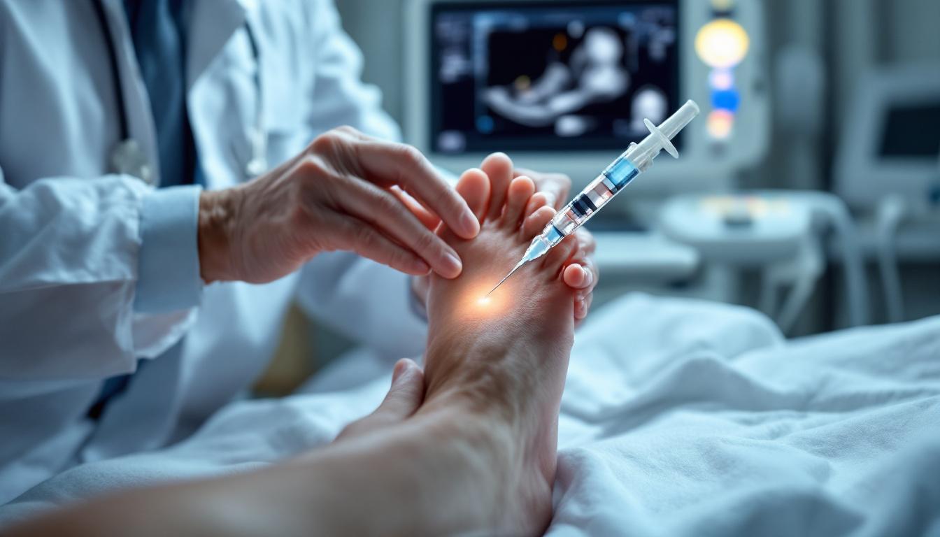
PRP for Morton's neuroma is delivered as a perineural injection, placed under ultrasound guidance in the intermetatarsal space, adjacent to the affected digital nerve. Ultrasound guidance is essential: the intermetatarsal space is small, the neuroma is compact, and precise perineural delivery is required for efficacy. The injection bathes the nerve and surrounding tissue in growth factors, aiming to reduce fibrosis, modulate pain signaling, and support the biological environment for nerve recovery.
Ultrasound-guided PRP injection for Morton's neuroma
A 2020 prospective study examining ultrasound-guided PRP injection for Morton's neuroma2 reported that a majority of patients achieved meaningful reduction in VAS pain scores and improved functional outcomes at 6-month follow-up. No serious adverse events were reported. The authors noted that patient selection, particularly avoiding cases with very large neuromas (>5mm), was important for treatment success.
The honest summary: the evidence for PRP in Morton's neuroma is early-stage and based on small trials. The comparison studies suggest PRP produces comparable short-term results to corticosteroid with potentially superior durability at 12 months, a meaningful advantage given the tendency of cortisone to require repeat injections. However, large randomized controlled trials are lacking, and PRP has not been validated in high-quality studies against alcohol sclerosing injections, which are also used for this condition. PRP is a reasonable second-line injectable option for patients with chronic Morton's neuroma who have had limited benefit from cortisone.
PRP vs. Cortisone for Morton's Neuroma
Corticosteroid injections remain first-line for Morton's neuroma and typically produce rapid relief within 1–2 weeks. The limitations are well-documented: benefit tends to fade by 3–6 months, and repeated cortisone injections into the forefoot carry specific risks, including plantar fat pad atrophy, skin and soft tissue changes, and potential for weakening of surrounding soft tissue structures. The fat pad provides critical cushioning under the metatarsal heads; its atrophy can create a new source of chronic forefoot pain.
PRP does not carry these atrophic side effects. For patients who have had 2–3 cortisone injections with declining durability or who are concerned about cumulative steroid exposure in the forefoot, PRP offers a biologically regenerative alternative. The onset is slower, improvement typically develops over 4–8 weeks, but the response may be more sustained based on the available comparison data. PRP is also considered before escalating to alcohol sclerosing injections or neurectomy.
Who Is a Good Candidate?
PRP is most appropriate for patients with ultrasound-confirmed Morton's neuroma (typically 5mm or smaller) who have not achieved lasting relief from footwear modifications, orthotics, and at least one to two cortisone injections. It is also reasonable for patients who want to minimize steroid exposure in the forefoot, or as an alternative to alcohol sclerosing injections for those who prefer a regenerative approach. The best candidates have failed conservative care but are not yet ready for surgical neurectomy.
Larger neuromas (over 6–8mm on ultrasound) tend to respond less well to any injection therapy and are more likely to require surgical management. Patients with bilateral or multiple interspace neuromas, or those with associated metatarsalgia from structural foot deformity, should have these contributing factors addressed as part of the treatment plan.
What to Expect
Morton's neuroma PRP is administered as an ultrasound-guided perineural injection in the intermetatarsal space, an outpatient procedure taking approximately 20–30 minutes. The ultrasound allows direct visualization of the neuroma and real-time guidance of the needle to the perineural target zone. Some forefoot soreness is expected for 2–5 days after the injection.
Tight footwear and high heels should be avoided for 2 weeks post-injection. Most patients can return to light walking the same day. Meaningful improvement typically begins at 4–8 weeks. One injection is typical; some practitioners offer a second injection at 6–8 weeks for partial responders. Concurrent footwear optimization and metatarsal pad use is important to reduce ongoing nerve compression.
1. Mahindra P, Yamin M, Selhi HS, Singla S, Soni A. Chronic Plantar Fasciitis: Effect of Platelet-Rich Plasma, Corticosteroid, and Placebo. Orthopedics. 2016;39(2):e285-9. doi:10.3928/01477447-20160222-01. PMID: 26757531.
2. Malahias MA, et al. Platelet-Rich Plasma Ultrasound-Guided Injection in the Treatment of Morton's Neuroma: A Case Series Study. J Foot Ankle Surg. 2020;59(2):287-292. PMID: 32019697.
This content is for educational purposes only and does not constitute medical advice. Consult a qualified healthcare provider before starting any treatment.