Explore how PRP injections can treat patellar tendinitis. Learn about platelet-rich plasma therapy benefits, recovery, and results for patellar tendinitis.
Patellar tendinopathy, commonly called jumper's knee, develops when repetitive loading causes degenerative breakdown of the tendon connecting your kneecap to your shin bone. Unlike true inflammation, the underlying pathology involves disorganized collagen fibers and arrested tissue repair. Platelet-rich plasma (PRP) therapy addresses this root cause by delivering a concentrated mix of growth factors directly into the degenerated tendon, reactivating the regenerative process that chronic overuse has disrupted.
PRP treatment for patellar tendinopathy
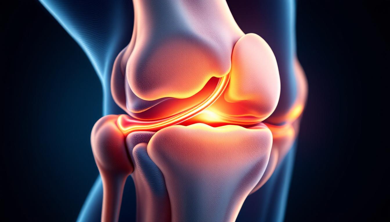
What Is Patellar Tendinitis?
The patellar tendon connects the bottom of the kneecap (patella) to the top of the tibia, transmitting the powerful contractile force of the quadriceps into knee extension. It is subjected to enormous repetitive loads during jumping, sprinting, and rapid deceleration. When cumulative stress outpaces the tendon's capacity for self-repair, the collagen matrix breaks down, a degenerative process characterized histologically by disorganized collagen, fibrocyte proliferation, and the absence of classic inflammatory infiltrates. Modern orthopedic literature favors the term patellar tendinopathy over patellar tendinitis for this reason.
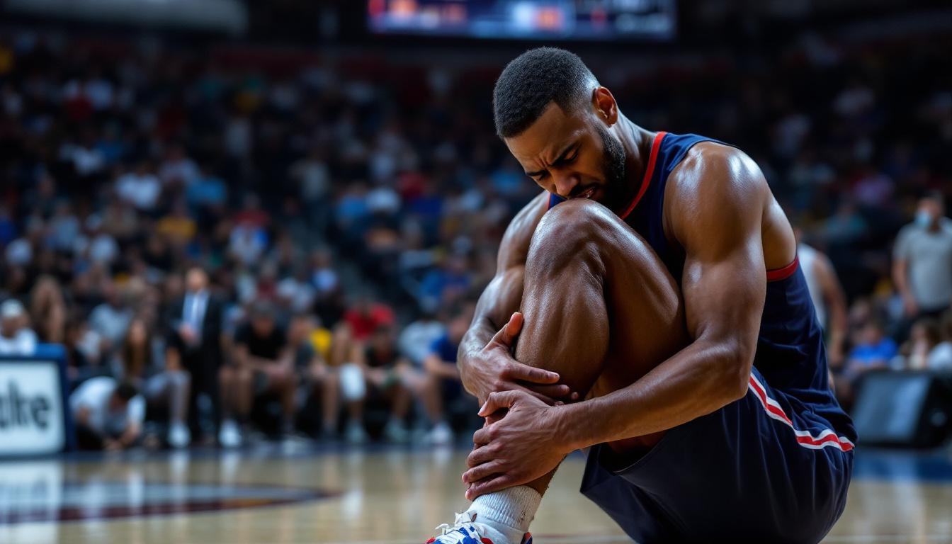
The condition is most prevalent among athletes in jumping sports, basketball, volleyball, and track, but it also affects recreational runners, cyclists, and active adults. The hallmark symptom is focal pain at the inferior pole of the patella that worsens with loading activities. In chronic cases, pain may persist at rest and significantly limit sports participation and daily function.
Application
Patient Profile
Evidence Level
Typical Protocol
Best Candidates
Chronic Patellar Tendinopathy
Jumper's knee symptoms lasting 3+ months, failed eccentric loading program and NSAIDs
Level I–II (multiple RCTs supporting PRP over placebo and cortisone)
1–3 ultrasound-guided injections at 4–6 week intervals with structured eccentric rehab
Athletes with mid-tendon pathology on ultrasound, no prior cortisone within 3 months
Partial Patellar Tendon Tear
MRI-confirmed partial thickness tear under 50% cross-section in high-load athletes
Level II–III (cohort studies, case series)
2–3 injections with progressive loading protocol and load management
Platelet-rich plasma is derived from the patient's own blood through centrifugation, concentrating platelets and the bioactive proteins they carry. Key growth factors in PRP, including platelet-derived growth factor (PDGF), transforming growth factor-beta (TGF-β), insulin-like growth factor (IGF-1), and vascular endothelial growth factor (VEGF), are essential mediators of tendon repair and collagen remodeling.
Under ultrasound guidance, PRP is injected directly into the area of degenerative change at the inferior patellar pole. The concentrated growth factors activate quiescent tenocytes, stimulate new collagen synthesis, promote angiogenesis, and help reorganize the disorganized extracellular matrix. This mechanism is fundamentally different from corticosteroid injection, which temporarily suppresses tissue activity but does nothing to restore the structural integrity of the tendon.
Most treatment protocols use one to three PRP injections spaced four to six weeks apart, combined with a structured eccentric-loading rehabilitation program. Evidence suggests that progressive mechanical loading during recovery, rather than complete rest, helps organize newly deposited collagen along functional stress lines, improving both strength and durability.
Ultrasound-guided PRP injection for patellar tendon degeneration
What the Research Shows
Clinical evidence for PRP in patellar tendinopathy has grown substantially, with randomized controlled trials and systematic reviews consistently demonstrating meaningful improvements in pain and function scores compared to control interventions.
A double-blind randomized controlled trial compared leukocyte-rich PRP to dry needling in patients with chronic patellar tendinopathy. The PRP group demonstrated significantly greater improvements in the Victorian Institute of Sport Assessment-Patellar (VISA-P) score at 12-month follow-up, supporting the conclusion that growth factor delivery provides a durable regenerative advantage over mechanical stimulation alone. Dragoo et al., Am J Sports Med 2014[1]
A systematic review of injection therapies for patellar tendinopathy found that PRP produced superior outcomes compared to corticosteroid injections at medium- and long-term follow-up, with the anabolic regenerative mechanism of PRP cited as the key driver of its sustained efficacy. Filardo et al., Int Orthop 2010[2]
PRP vs. Cortisone for Patellar Tendinopathy
Corticosteroid injections have historically been used to manage tendon pain, but current evidence raises significant concerns about their long-term use in tendinopathy. Cortisone is a catabolic agent: while it reliably reduces pain in the short term, it does so by suppressing cellular activity in a tissue that is already failing to repair itself. Multiple studies have linked repeated corticosteroid injections to tendon weakening, collagen fiber disruption, and, in some cases, increased risk of tendon rupture.
PRP works through the opposite mechanism. By delivering anabolic growth factors to the site of degeneration, it stimulates the repair and remodeling processes that tendinopathy has arrested. Clinical trials comparing the two approaches consistently show that PRP produces inferior short-term pain relief but significantly better outcomes at six to twelve months, consistent with its regenerative, rather than palliative, mode of action. For patients with chronic patellar tendinopathy who have failed physical therapy, PRP represents a more durable therapeutic strategy.
Who Is a Good Candidate?
PRP is most appropriate for patients with confirmed patellar tendinopathy, typically diagnosed by clinical examination and ultrasound or MRI, who have not achieved adequate relief after three to six months of conservative management, including eccentric strengthening, load modification, and physical therapy. It is also considered for competitive athletes who need a faster return to sport or cannot tolerate extended rest periods.
Patients with active infection, bleeding disorders, or systemic inflammatory conditions may not be candidates. A consultation with a sports medicine or orthopedic specialist is the appropriate first step to determine whether PRP is right for your presentation.
What to Expect
Following injection, mild soreness at the injection site is common and typically resolves within three to five days. Most patients begin a guided rehabilitation protocol within one week. Meaningful improvement in pain and function is usually observed between six and twelve weeks, with continued gains through six months. Athletes typically require four to eight weeks before returning to full training loads, depending on severity of degeneration and individual healing response.
1. Dragoo JL, Wasterlain AS, Braun HJ, Nead KT. Platelet-rich plasma as a treatment for patellar tendinopathy: a double-blind, randomized controlled trial. Am J Sports Med. 2014;42(3):610-618. PMID 24136862
2. Filardo G, Kon E, Villa SD, Vincentelli F, Fornasari PM, Marcacci M. Use of platelet-rich plasma for the treatment of refractory jumper's knee. Int Orthop. 2010;34(6):909-915. PMID 19728039
This content is intended for informational purposes only and does not constitute medical advice. Consult a qualified healthcare provider before beginning any treatment program.
Frequently Asked Questions
Find a Patellar Tendinitis PRP Specialist Near You
Work with a sports medicine provider experienced in ultrasound-guided PRP for tendon injuries.